Recently, the incident of "a patient was forgotten for 6 hours while undergoing MRI" has aroused widespread public concern about radiation problems in imaging examinations. Tongji Hospital has suspended the medical staff involved, conducted a comprehensive examination of the patient, and negotiated compensation matters. CCTV News interviewed experts to explain in detail the radiation differences of common imaging examinations such as MRI, B-ultrasound, CT, and
Experts have made it clear that there is no ionizing radiation in MRI and B-ultrasound examinations, so there is no need to worry about radiation damage.
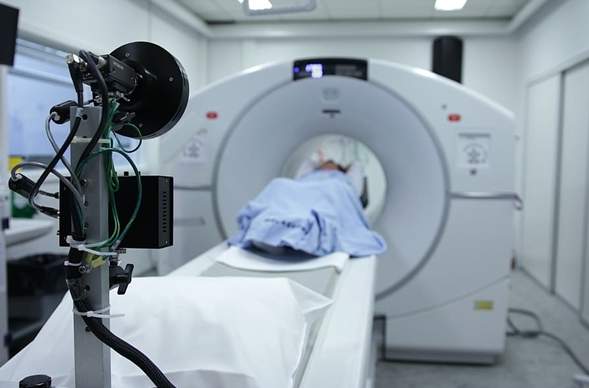
Among them, nuclear magnetic resonance uses strong magnetic fields and radio waves to adjust the magnetic direction imaging of hydrogen atoms. It is an important method for diagnosing soft tissue diseases such as the nervous system and musculoskeletal system. Even if the patient is in the examination room for a long time, he will only endure the psychological pressure of the claustrophobic space, machine noise and physical fatigue, without radiation risks.
However, it is necessary to guard against the hidden dangers caused by strong magnetic fields: People with ferromagnetic metal implants such as pacemakers and cochlear implants are strictly prohibited from examination. Metal objects are not allowed to be brought into the examination room. If tattoo pigments contain metal, the doctor must be informed in advance.
B-ultrasound uses ultrasonic echo to produce real-time imaging, which is non-invasive, convenient and radiation-free. It is the preferred examination method for pregnant women and children, and is suitable for the examination of liver, gallbladder, pancreas, thyroid, breast and other organs.
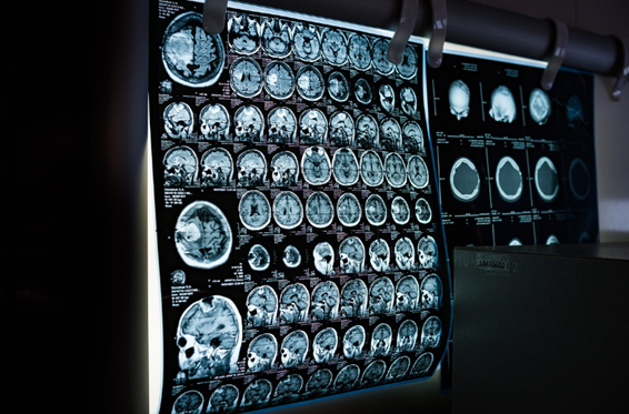
Nuclear medicine examinations such as X-ray, CT, and PET-CT all involve ionizing radiation that needs to be guarded against. These examinations are precise special examinations that can clearly determine subtle lesions in the lungs and brain, but there is a significant difference in radiation dose.
-X-ray photography uses X-rays to form a flat image and is often used to examine fractures and lung inflammation. The radiation dose is extremely low. One examination is equivalent to 10 days of natural background radiation.
-CT reconstructs tomographic images through X-ray multi-layer scanning, which can display subtle lesions in various organs of the body, such as early stage lung cancer, cerebral hemorrhage, etc. The amount of information far exceeds that of X-rays, but the radiation dose is higher. The statement "one CT is equivalent to taking hundreds of X-rays" is basically accurate. The radiation dose of one chest CT is equivalent to 2-3 years of natural background radiation.
-PET-CT requires the injection of radioactive tracers and combines functional metabolism and anatomical structure imaging. It is an important means for early diagnosis, staging and efficacy evaluation of tumors.
Experts emphasize that the risk of a single necessary examination with radiation such as CT is extremely low, and the public does not need to stop eating due to choking. For example, low-dose spiral CT screening for people at high risk of lung cancer once a year is an important early screening method.
For special groups such as pregnant women, couples preparing for pregnancy, and infants and young children, experts give clear recommendations for examination selection:
-B-ultrasound is absolutely safe for pregnant women and fetuses, and is the first choice for examination.
- Magnetic resonance imaging also has no radiation, but it is not recommended in early pregnancy. Infants and young children may need sedation and cooperation.
- Pregnant women and couples preparing for pregnancy should be careful when choosing X-rays. When examination is necessary, it can be carried out under the protection of lead clothing. For those who are preparing for pregnancy, it is recommended to wait 3-6 months before preparing for pregnancy.
-Strictly limit CT and PET-CT.