Microplastics are almost everywhere, with tiny plastic particles found in everything from water samples to tissue biopsies to autopsy specimens. Now, for the first time, scientists have "seen" the existence of these microplastics and their movement paths in vivo.A research team from University College London (UCL), the University of Birmingham and Kingston University used a laser imaging technology to image and track microplastics in deep tissues in mice without surgery, providing a new perspective on how microplastics migrate, accumulate and have long-term health effects in the body.
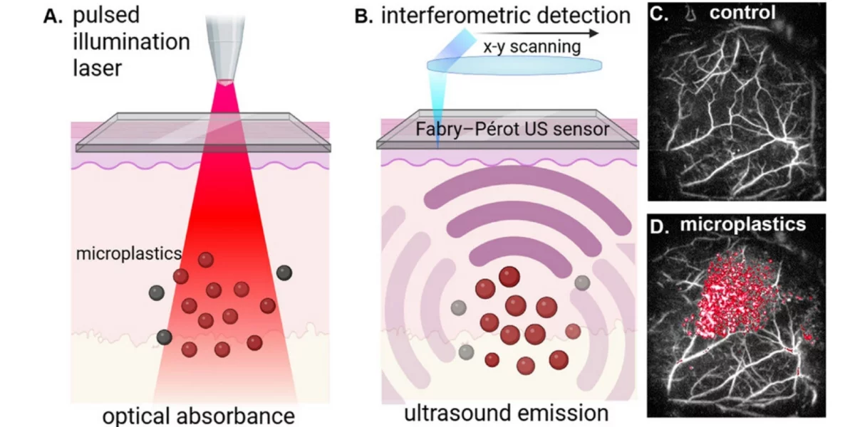
The technology, called "photoacoustic imaging," emits short pulses of laser light into tissues, causing microplastic particles to absorb light energy and generate high-frequency sound waves. The ultrasonic detector then receives the signal and reconstructs the image, thereby mapping the distribution of microplastics in the body. Different microplastics have their own unique light absorption fingerprints, allowing researchers to distinguish and target the location of these particles in complex tissue environments. In the experiment, the researchers injected mice with about 0.5 milligrams of microplastic—"roughly the visual equivalent of a handful of very fine grains of salt"—and then monitored its migration through living tissue over an extended period of time.
Stephen Patrick, a lecturer in medical imaging at University College London, said that the team can accurately track the movement of microplastics on a time scale of months instead of days, closer to their actual behavior in the human body. Research shows that this method can be used to obtain more detailed observations of where microplastics accumulate in the body, how long they stay, and whether they are involved in the occurrence of diseases in the brain, blood vessels and other organs. The basis of this imaging method is the pigments added to consumer plastic products for coloring purposes. These pigments provide identifiable signals for photoacoustic imaging.
Currently, black, gray, green and blue microplastics are the easiest to detect, so the research team selected common plastic items in daily life as sample sources, such as black ballpoint pen caps and green beverage bottle caps - previous studies have shown that bottle caps release microplastic particles during the tightening and unscrewing process. Patrick pointed out that if the typical distribution of microplastic colors in the human body can be grasped, the overall content can be more accurately estimated based on the "visible part." The technology's current estimates of the total amount of microplastics in the body are still conservative, but it has successfully identified several common types of microplastics, including polypropylene, which is widely used in food containers and coffee cups, and polyethylene, which is found in single-use plastic bags.
Concerns about the impact of microplastics on human health are rising. They have been found in the blood, organs and tissues, and have been linked to various health risks including cancer, myocardial infarction and reproductive problems. However, previous studies often relied on tissue analysis after biopsy or dissection, which had obvious limitations in the time dimension, making it difficult to truly present the dynamic process of long-term migration and accumulation of microplastics in vivo. At the same time, traditional chemical labeling methods may not only change the original behavior of microplastics, but also easily misjudge lipid substances in the body as microplastic signals.
Patrick said that in some existing methods, the high levels of "polyethylene" detected in brain tissue are likely to be fatty acid signals mistaken for microplastics. In contrast, body fat does not produce confounding signals at the currently used photoacoustic imaging bands, but the team still needs to confirm that other potential pigments do not cause similar interference. The new technology has been proven to be able to detect individual microplastic particles around 45 microns in size, which is smaller than the diameter of an average human hair.
For smaller particles, especially nanoplastics, current experiments have not been fully validated, but unpublished results show that detection is theoretically possible at concentrations as high as those reported in previous studies (on the low mg/ml scale). At lower concentrations, detection will be more challenging. Patrick believes that there is still room for significant improvement in technical accuracy by introducing more "techniques" and more complex image processing in the signal acquisition and processing links, because the current work only uses relatively simple system configurations and image processing solutions.
In future clinical studies, it would be ideal to cross-validate measurements obtained from photoacoustic imaging with other independent methods, such as using samples from patients who have undergone tissue resection for disease diagnosis or treatment. Such validation is considered a necessary step before the technology can be used independently in the clinic to assess microplastic burden in patients. Follow-up research directions may include: systematically teasing out the transport, retention, and clearance mechanisms of microplastics in the body, exploring how these processes change with particle size, shape, and underlying disease conditions, and further analyzing their association with conditions such as vascular disease and liver cirrhosis.