
Abstract:
A hospital in London, England, recently announced that a medical team successfully completed a brain tumor resection for the first time with the assistance of an artificial intelligence system. During the operation, AI will analyze the real-time video of the surgical field and color-mark key nerves and blood vessels to help doctors avoid structures that may cause serious damage when removing tumors.
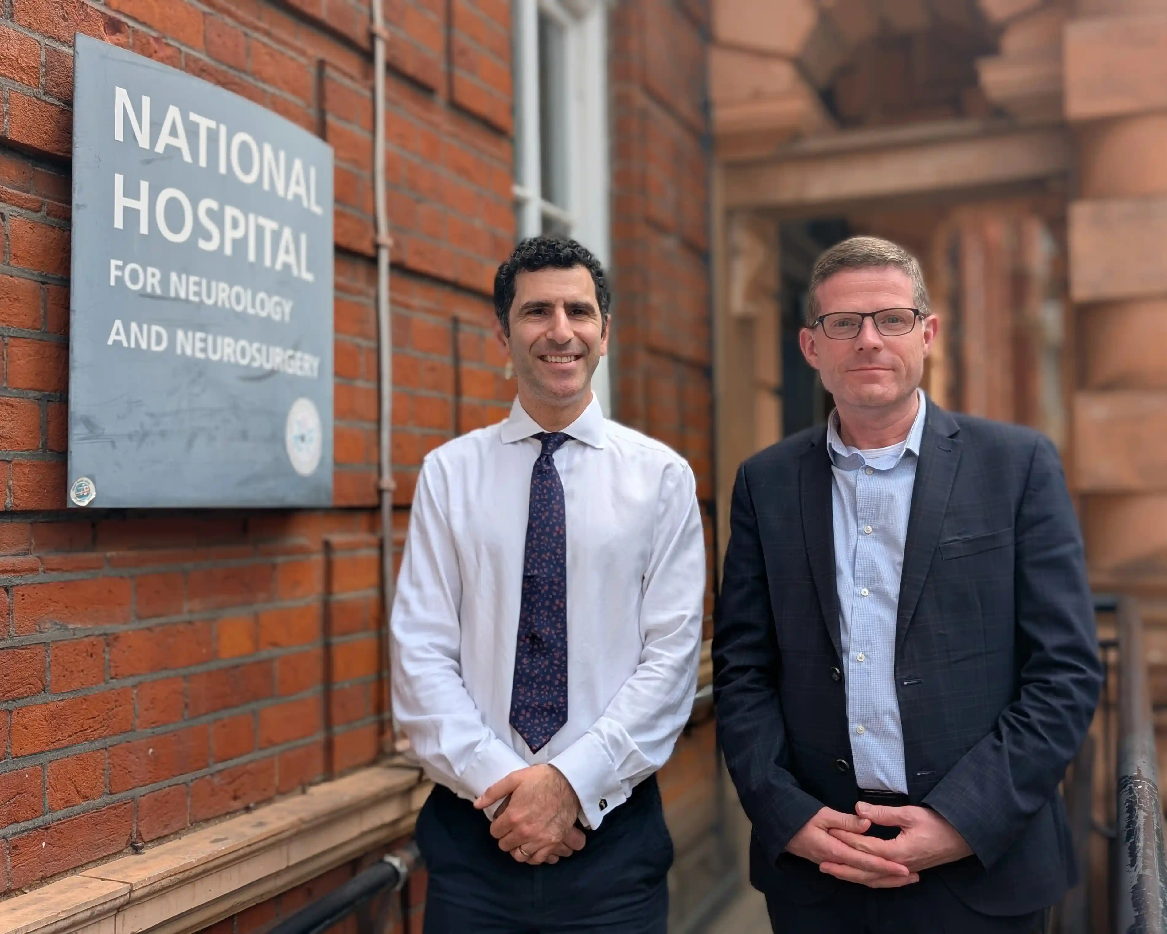
The operation was performed in May this year at the National Hospital for Neurology and Neurosurgery in London, which is part of the University College London Hospitals NHS Foundation Trust and is part of the National Health Service. The hospital said this was the first successful operation to use AI to assist in the removal of a brain tumor, but the news was not announced to the public until after the patient recovered on Thursday.
The person who underwent the operation was 48-year-old Bedfordshire resident Rhys Hibbert. He had a tumor about 11 mm in diameter on his pituitary gland. Nerves responsible for visual function and important blood vessels are distributed near the pituitary gland, so there is very little room for surgical error. Health officials point out that when operating in this area, even a deviation of just 1 millimeter can lead to blindness, stroke, and even death.
Previously, Hibbert was diagnosed with the tumor in 2024, and doctors initially used non-surgical methods to control the disease. But over time, he developed severe hormone imbalances and worsened vision problems. The hospital said that if he did not undergo the surgery, he might eventually become blind.
In this operation, the AI system analyzed the surgical images in real time and color-coded the anatomical structures that needed to be protected, allowing the surgical team to more clearly identify nerves, major blood vessels, and other tissues. The technology has been used in hospital research projects in the past, but has never before been used in actual patients during surgery.
After the surgery, Hibbert's vision improved rapidly. He said that he could clearly see everything in the room after waking up. Within a week after surgery, he was no longer wearing glasses or walking with a cane and was back at work as a customer service manager. He described the feeling after recovery as "gaining a 360-degree view."
The technical development of the system is led by Sophia Barno, associate professor of robotics and artificial intelligence at University College London. She said that by learning hundreds of surgical videos, the software has been exposed to and analyzed a large number of different cases, so it can identify the interactions between key anatomical structures, surgical instruments and tissues during the operation, providing additional support for doctors to handle difficult and delicate operations.
The surgery was funded by the National Institute for Health and Care Research and was carried out as part of a clinical trial. Mike Lewis, scientific director of innovation affairs at the agency, said this case demonstrates the potential of advanced AI to assist doctors and improve patient care in the surgical field.
Hibbert pointed out that the National Hospital of Neurology and Neurosurgery in London was founded in 1859 and was the first hospital in the world to specialize in neurosurgery diagnosis and treatment. Now, this hospital has completed its first AI-assisted neurosurgery, which he believes has special symbolic significance.
Comments