
Abstract:
A new study by U.S. and Australian researchers adds new evidence to the ongoing debate over whether total joint replacement increases the risk of Alzheimer's disease. The research team analyzed about 700 autopsy human brain samples and found that materials from joint replacements can indeed migrate into neural tissue and may increase the content of proteins associated with Alzheimer's disease. However, the researchers did not find that the particles affected brain function.
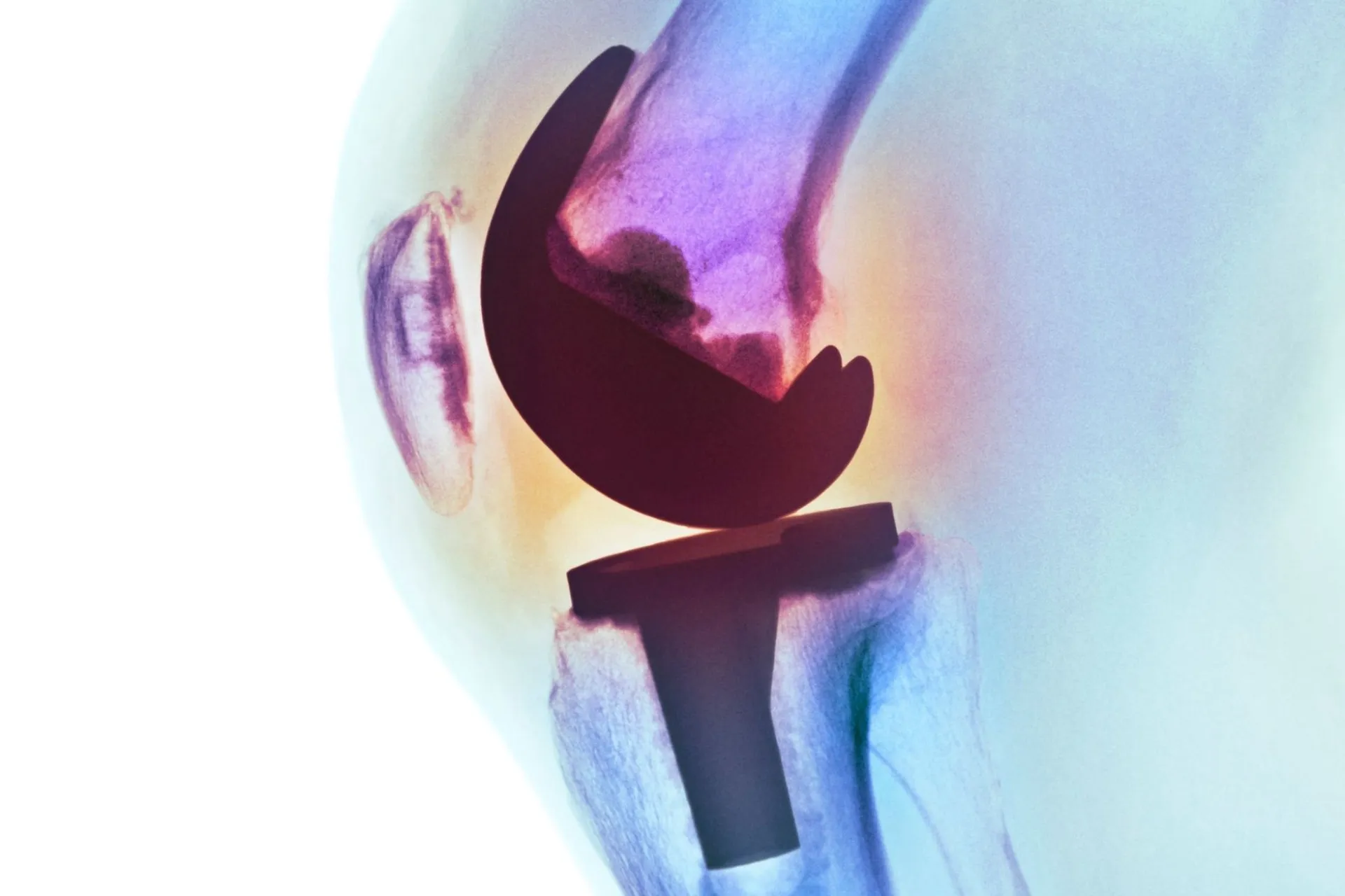
This is good news for people who have received joint implants or are preparing for surgery. However, the researchers also pointed out that the potential impact of tiny metal debris on cognitive health cannot be ruled out.
"We've known for years that microparticles can be deposited in the tissue around joints, but this is the first study to examine whether microparticles from joint replacements can be deposited in the brain," said Robin Pulzar, director of the Division of Implant Materials Analysis in the Department of Orthopedic Surgery at Rush University.
More than 1.5 million patients with osteoarthritis in the United States undergo total joint replacement surgery each year. Meanwhile, approximately 7 million people in the United States are living with Alzheimer's disease. Since both diseases are more common in older adults, it is not surprising that there is a large overlap between the two.
However, whether this association is entirely due to age has long remained an open question.
Theoretically, submicron particles generated by implants over the course of years of wear and tear may enter the brain, thereby increasing the risk of neurodegeneration. Studies have shown that debris generated by implants can induce local immune responses and can be detected in the blood and other organs.
To determine whether implants containing cobalt-chromium-molybdenum alloys and titanium-aluminum-vanadium alloys could cross the blood-brain barrier, researchers collected autopsy samples from deceased volunteers from Rush University's Memory and Aging Project.
Of the 885 participants, 701 brain samples were ultimately included in the metal content analysis. Among them, 229 volunteers had undergone shoulder, knee or hip replacement surgery; the other 472 had not undergone joint replacement and were therefore used as a control group.
Spectral analysis showed a correlation between cobalt concentrations in four areas of the brain and the surgery in people who had undergone total hip replacements. The relevant content in brain tissue samples from these people was about 8.9% higher. Electron microscopy also found cobalt particles in the samples.
The researchers also found that cobalt levels were associated with small increases in amyloid beta levels in the brain's inferior temporal cortex. In contrast, titanium content was inversely correlated with amyloid beta burden.
The key is that neither the presence of cobalt nor titanium was associated with changes in cognitive ability.
The study results overall further support the safety of joint replacement surgery. Each year, these surgeries help millions of people around the world regain mobility and significantly improve their quality of life.
"Total joint replacement remains one of the most successful and life-changing interventions in modern medicine," Pulzar said. "But this study does suggest that particles from implant wear, particularly from certain hip replacement implants that experience accelerated wear, may migrate beyond the joint and warrant further study."
The research was published in Acta Biomaterials.
Comments