
Abstract:
MIT engineers have developed a thin underwater robot powered by living skeletal muscle cells. The device does not have a traditional motor. The main body is two independently controllable gel fins, each about 15 mm long, 7 mm wide, and 0.5 mm thick. The surface is covered with a thin layer of muscle cells less than 15 microns thick. The research team allowed mouse skeletal muscle cells to express photosensitive ion channels that can respond to blue light. After irradiating blue light with a wavelength of about 470 nanometers, the cells contract and drive the gel fins to flap, pushing the robot to move in the water.
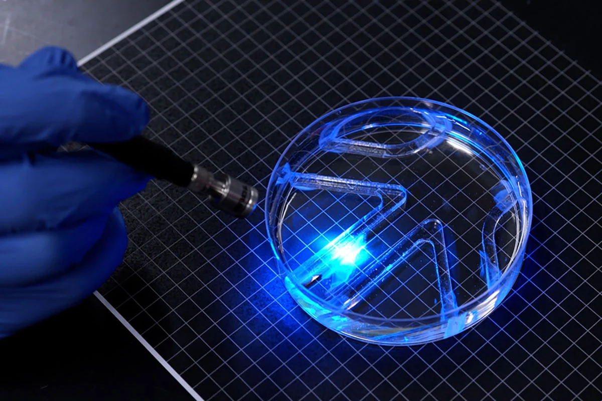
The research results were published in "Advanced Functional Materials". The team made a scaffold from methacrylated gelatin called GelMA and imprinted grooves on the surface to guide the muscle cells to align in the same direction. Compared with the previously used fibrin gel, the GelMA scaffold is better able to withstand muscle contraction and is less likely to shrink with cell movements. Experiments also found that square grooves at the bottom are more conducive to muscle cell arrangement, fusion into fibers, and coordinated force generation than arc-shaped grooves. The researchers also "trained" the muscle tissue through repeated flash stimulation to enhance its contraction displacement. Finally, two pieces of separately controlled muscle tissue form the left and right fins of the robot. Researchers can drive one side individually or drive both sides at the same time to change the direction and speed of travel.
In a petri dish tank, the researchers manually moved the light source to guide the robot through a simple maze. The maximum linear speed reported in the paper is about 4 times the fuselage length per minute, and the maximum rotation speed is about 1200 degrees per minute. That's far slower than an Olympic swimmer, but similar to a slow-moving bull shark, MIT said. The "navigation" here relies on external personnel to control the light source, and the robot does not yet have autonomous perception, decision-making or onboard control systems.
The paper also reports that the unit volume contraction force of the optimized two-dimensional muscle film is approximately 20 times that of three-dimensional muscle tissue; its functional maintenance time after being separated from fixed support has been improved from less than 10 minutes for the old fibrin structure to more than 30 days for the GelMA structure. This 30 days refers to the functional lifespan of the muscle membrane actuator without fixed support in the experiment, and does not mean that the robot can swim continuously for a month. Studies have shown that tissue displacement after light stimulation training is approximately 4 times that of the untrained control group.
This design aims to solve the problem of traditional biohybrid robots being large in size and requiring a large number of three-dimensional cultured muscle cells. The research team believes that living tissues that are soft, responsive to the environment and have certain self-healing capabilities may be used for tasks such as exploring fragile waters and environmental monitoring in the future. However, the research still relies on a single mouse muscle cell line, simple fins and external blue light stimulation; the team said that the next step will be to optimize the body structure to increase speed and explore the integration of micro-light sources and portable power supplies. Applications such as minimally invasive surgery are currently only long-term ideas and have not yet been verified in practice.
Comments