
Abstract:
A team from the Ecole Polytechnique Fédérale de Lausanne in Switzerland has developed a new fluorescence microscopy imaging method called SPIFFI, which can reconstruct super-resolution images from a single camera exposure and help researchers observe rapid changes in living cell structures such as mitochondria and microtubules. The research was published in the journal Nature Methods.
Traditional partial super-resolution microscopy requires continuous shooting of hundreds or even thousands of images, and then uses fluorescence changes to reconstruct details; when cells are active, structures will move or change shape during shooting, resulting in blurry final images. SPIFFI instead uses the luminescence polarization information of fluorescent molecules: the optical system splits the fluorescence into four channels that are sensitive to different polarization directions, and then compares the images obtained from the same exposure to extract spatial details that are difficult to display with conventional wide-field microscopes.
In the experiment, the research team increased the imaging resolution by about 1.7 times with a single exposure, able to resolve structures of about 160 to 170 nanometers, and photographed the movement of mitochondria and microtubules in living cells, as well as processes such as the fusion and division of cell structures. Since each frame can generate a super-resolution image independently, consecutive frames can further form a living cell super-resolution video.
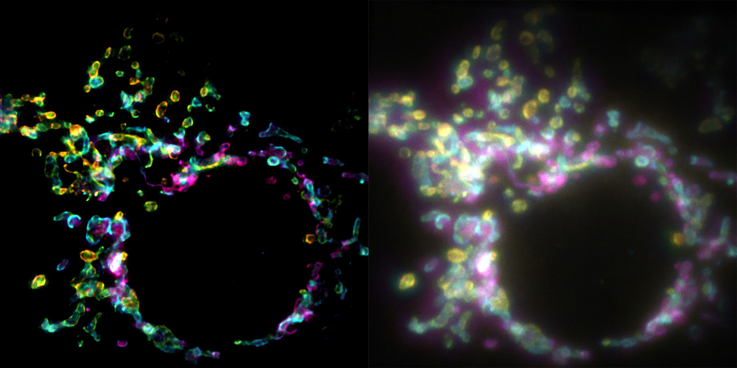
The researchers also combined the SPIFFI image with existing fluorescence fluctuation analysis methods and further improved the resolution to about 80 nanometers through post-processing; this value is not directly achieved by a single exposure. The team is currently trying to reduce the size of the device and combine it with three-dimensional microscopic imaging. Since the relevant optical components are expected to be integrated into existing fluorescence microscopes, this technology may be used in future research in cell biology, neuroscience, biophysics, and drug discovery.
Comments